
Aneurysm Rupture Treatment
ANEURYSM RUPTURE TREATMENT
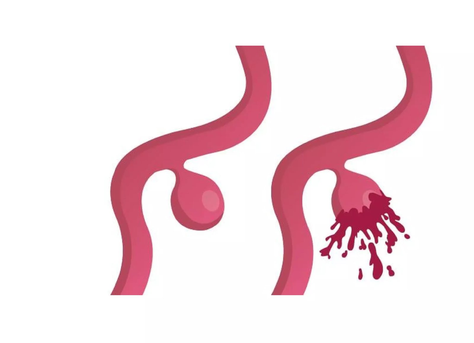
An intracranial aneurysm is a ballooning in the wall of an artery within the brain. The causes of aneurysms are not clear, but they can be congenital, genetics, due to high blood pressure, and certain medical conditions. The majority of aneurysms do not cause symptoms unless they rupture. If ruptured, it causes subarachnoid hemorrhage or intracerebral (within the substance of the brain) hemorrhage.
It is a medical emergency requiring immediate attention. Prompt diagnosis and intervention are vital to patient survival and neurological preservation.
Diagnosis of a ruptured aneurysm is clinical (sudden explosive headache and neck stiffness) and is confirmed by the presence of blood in cerebrospinal fluid (CSF) via lumbar puncture and on an emergency CT Scan of the brain.
Investigation: The precise anatomical presence, architecture, and morphology of a ruptured aneurysm is confirmed by digital subtraction angiography (DSA) or CT angiography (CTA) of cerebral vessels.
Treatment: Treatment of aneurysm aims to obliterate the balloon projecting out from the parent artery. This is achieved by two primary types of procedures:
CRITICAL EMERGENCY: THE FIRST 24 TO 72 HOURS
A ruptured brain aneurysm carries an immediate risk of catastrophic re-bleeding. Ultra-urgent surgical clipping or endovascular coiling permanently secures the aneurysm and protects against fatal vasospasm.
Symptoms of Ruptured Aneurysm
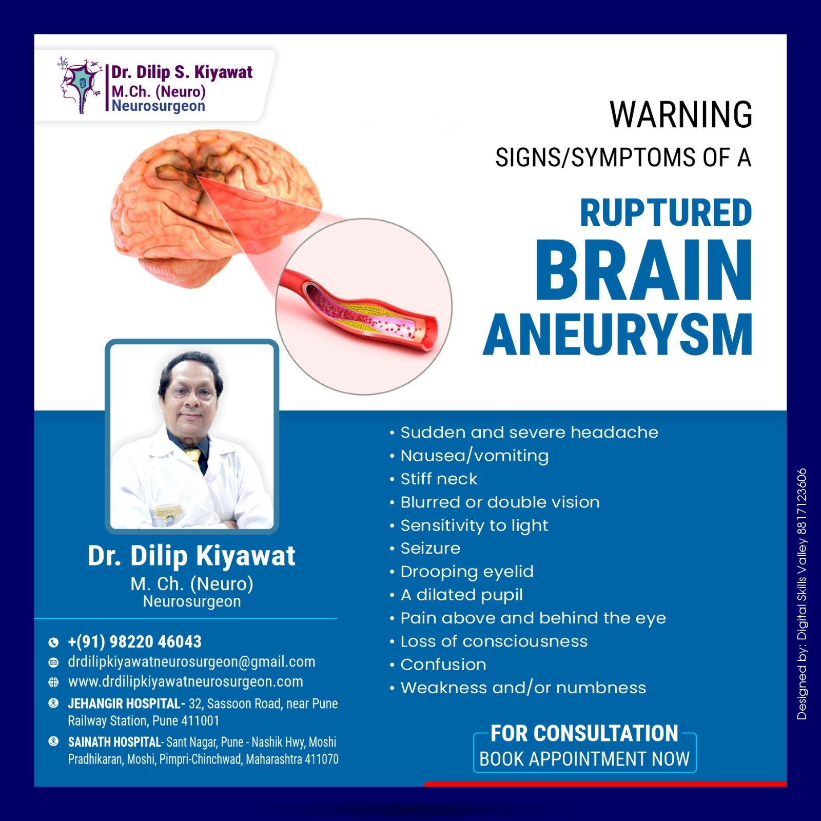
Recognizing these hallmark warning signs enables immediate life-saving intervention:
Sudden, Severe Headache
Often described by patients as “the worst headache of my life” (thunderclap headache), peaking within seconds to minutes of onset.
Nausea & Vomiting
Triggered by acute elevation of intracranial pressure and severe chemical irritation of basal meninges by extravasated blood.
Stiff Neck (Meningismus)
Pain and stiffness in the neck are noticed when the neck is bent forward, caused by subarachnoid blood pooling around the cervical spinal cord.
Sensitivity to Light (Photophobia)
Intense eye discomfort or inability to tolerate normal indoor and outdoor illumination due to meningeal irritation.
Blurred or Double Vision (Diplopia)
Compression or ischemia of cranial nerves (particularly the third cranial nerve / oculomotor palsy) resulting in pupillary dilation and visual disturbance.
Loss of Consciousness & Coma
In severe cases, a ruptured aneurysm leads to rapid loss of consciousness, motor seizures, confusion, or progression into coma requiring immediate ICU ventilatory support.
Clipping vs. Endovascular Coiling
The choice between these procedures depends on factors such as the aneurysm’s location, neck morphology, size, and the patient’s overall health:
1. Microsurgical Clipping
After opening the skull (craniotomy) under high-power surgical magnification, the arachnoid cisterns are dissected to expose the parent artery and aneurysm neck. A small, biocompatible titanium metal clip is placed across the neck of the aneurysm to isolate it from the blood circulation, preventing further rupture permanently.
- Complete and permanent exclusion with near-zero recurrence
- Ideal for wide-necked, middle cerebral artery (MCA), and complex bifurcations
- Allows direct evacuation of concurrent intracerebral hematomas
2. Endovascular Coiling
In this minimally invasive technique, a microcatheter is threaded through the femoral artery in the groin under fluoroscopic guidance to reach the cerebral aneurysm site. Small, flexible platinum coils are then placed sequentially within the aneurysm dome, promoting thrombotic occlusion and sealing it off from circulation.
- No cranial incision or bone flap removal required
- Favorable for posterior circulation (basilar, vertebral) and elderly patients
- Shorter hospital stay and faster post-procedural mobilization
Causes & Etiology
Aneurysms are often caused by a weakening of the blood vessel wall, which may be congenital, genetic, or acquired over time due to hypertension and hemodynamic stress.
Symptoms
Sudden, Severe Headache. A rapid-onset, intense headache is a common symptom accompanied by stiff neck, vomiting, photophobia, and visual impairment.
Emergency Treatment
An aneurysm rupture is a medical emergency. Immediate medical attention is crucial to stabilize cerebral perfusion and achieve definitive aneurysm exclusion.
Rehabilitation
Ongoing support for both the individual and their caregivers is crucial for the rehabilitation process, cognitive re-training, speech therapy, and blood pressure monitoring.
Clinical Radiographs & Aneurysm Case Studies
Authentic diagnostic neuroimaging and surgical records from Dr. Dilip Kiyawat's cerebrovascular archives:
 Zoom
Zoom
Brain Aneurysm Rupture Scaled
High-resolution diagnostic angiogram revealing acute saccular outpouching of the cerebral arterial wall with localized contrast extravasation and subarachnoid spread.
 Zoom
Zoom
Coiling of Aneurysm
Intra-procedural fluoroscopic documentation confirming compact deployment of detachable platinum microcoils, completely sealing the aneurysm neck from circulation.
 Zoom
Zoom
Ruptured Brain Aneurysm Case
Emergency CT brain demonstrating extensive hyperdense subarachnoid blood layering within the suprasellar and perimesencephalic cisterns with acute hydrocephalus risk.
For emergency aneurysm evaluation and 24/7 neurovascular consultation, reach Dr. Dilip Kiyawat at
-
Phone / 24/7 Emergency +91 98220 46043
-
Website drdilipkiyawatneurosurgeon.com