
Hydrocephalus
HYDROCEPHALUS
Hydrocephalus is a condition characterized by an abnormal accumulation of cerebrospinal fluid (CSF) in the brain’s ventricles, leading to increased intracranial pressure. This can result from various causes, including congenital conditions, infections, or head injuries. Symptoms may include headaches, nausea, impaired vision, and cognitive difficulties. In infants, progressive enlargement of head size is commonly noted.
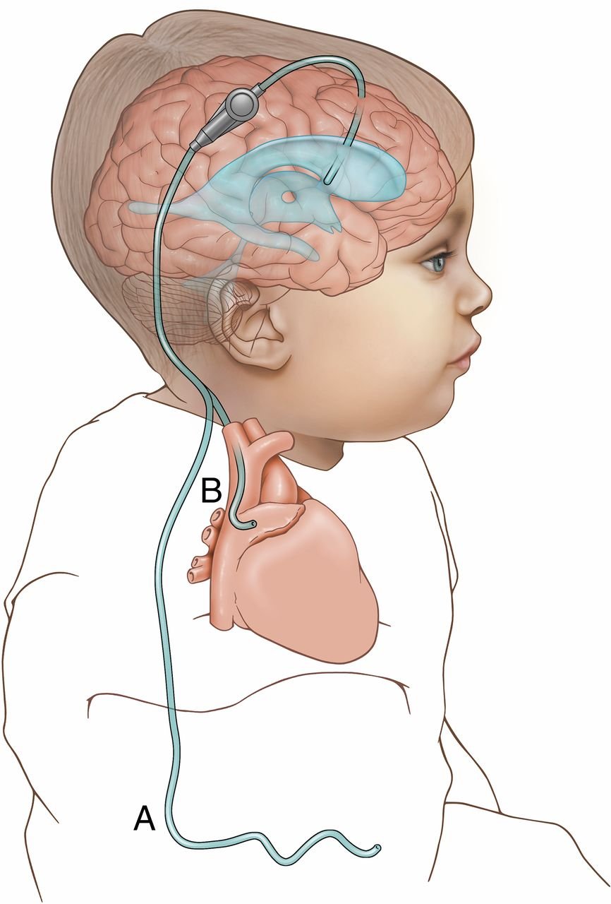
Treatment typically involves surgical intervention, such as the insertion of a shunt (ventriculoperitoneal shunt) to redirect excess CSF from the ventricles to the peritoneal cavity in the abdomen, relieving intracranial pressure.
Regular monitoring and follow-up are essential to address potential complications and ensure the optimal functioning of the shunt system, contributing to improved quality of life for individuals with hydrocephalus.
CSF CIRCULATION & VENTRICULAR RESTORATION
Normalizing intracranial pressure, safeguarding cognitive development, and preserving cortical mantle thickness through precision VP shunt placement and Endoscopic Third Ventriculostomy (ETV).
Causes
Blockage that prevents the normal flow of cerebrospinal fluid can lead to hydrocephalus. This obstruction can occur within the ventricles or in the passages connecting them (aqueductal stenosis, tumors, or arachnoid webs).
Symptoms
Downward deviation of the eyes (‘sunsetting sign’), vomiting, irritability, sleepiness, headache, nausea, blurred or double vision, and rapid head circumference enlargement in infants.
Treatment
The most common treatment for hydrocephalus involves the surgical placement of a shunt (Ventriculoperitoneal / VP Shunt) with adjustable valves or minimally invasive neuro-endoscopic surgery.
Rehabilitation & Care
Some cases may be managed with medication to reduce CSF production temporarily. Comprehensive developmental monitoring, pediatric neuro-rehabilitation, and regular valve checks ensure lasting recovery.
Breakthrough Treatment Shows Promise in Hydrocephalus
Contemporary neurosurgical techniques for pediatric, adult, and normal pressure hydrocephalus:
Post-Infectious Hydrocephalus
Infections affecting the central nervous system, such as bacterial or tuberculous meningitis, can lead to severe inflammation, arachnoid adhesions, and subsequent blockage of normal CSF pathways.
Medical Management
Some temporary or borderline cases may be managed with medication (such as acetazolamide or furosemide) to reduce the production of cerebrospinal fluid under close neuro-imaging surveillance.
Endoscopic Third Ventriculostomy (ETV)
In selected cases—especially in non-communicating hydrocephalus, older children, and adults—a minimally invasive endoscopic procedure creates a natural fenestration in the floor of the third ventricle, eliminating the need for permanent hardware.
Favorable Long-Term Prognosis
With appropriate and timely neurosurgical intervention, the prognosis for individuals with hydrocephalus is highly favorable, enabling children and adults to attain normal cognitive, motor, and intellectual milestones.
Advanced Surgical Solutions for Hydrocephalus
Dr. Dilip Kiyawat provides tailored interventions based on patient age, etiology, and CSF hydrodynamics:
Ventriculoperitoneal (VP) Shunt Surgery
Gold Standard ProcedureA specialized silicone catheter system is placed into the cerebral lateral ventricle through a small cranial burr hole. The catheter connects to a precision one-way differential or programmable valve and tunnels subcutaneously behind the ear, down the chest, and into the peritoneal cavity where excess fluid is absorbed naturally.
- Non-invasive magnetic valve programmability
- Integrated anti-siphon and gravitational flow control
- Lifelong durable CSF diversion with minimal recovery time
Endoscopic Third Ventriculostomy (ETV)
Hardware-Free EndoscopyUtilizing a high-definition rigid neuro-endoscope, Dr. Kiyawat navigates through the ventricle to the floor of the third ventricle (tuber cinereum). A delicate opening is made into the interpeduncular cistern, creating a physiological internal bypass that restores natural CSF circulation without an artificial implant.
- Completely foreign-body free — no hardware malfunction risks
- Ideal for obstructive aqueductal stenosis and tectal lesions
- Preserves native CSF absorption and pulsatile dynamics
Clinical Radiographs & Hydrocephalus Documentation
Authentic diagnostic neuro-imaging and procedural cases from Dr. Dilip Kiyawat's clinical archives:
 Zoom
Zoom
Hydrocephalus Ventriculomegaly
Diagnostic neuroimaging scan demonstrating severe symmetrical dilation of the cerebral ventricles with effacement of cortical sulci and elevated intracranial fluid volume.
 Zoom
Zoom
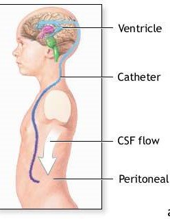
Ventriculoperitoneal Shunt Placement
Clinical surgical schematic and radiographic tracking of precision ventricular catheter insertion, reservoir placement, and distal peritoneal CSF diversion.
 Zoom
Zoom
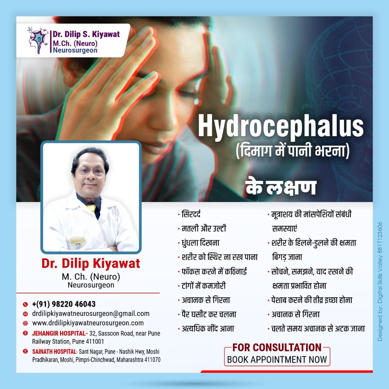
Symptoms of Hydrocephalus
Comprehensive clinical assessment mapping classical signs including sunsetting eyes sign, bulging fontanelle, projectile vomiting, and elevated intracranial pressure.
For hydrocephalus evaluation, second opinions, or shunt appointments, reach Dr. Dilip Kiyawat at
-
Phone / Clinic Line +91 98220 46043
-
Website drdilipkiyawatneurosurgeon.com