
Spina Bifida Surgery
SPINA BIFIDA SURGERY
Spina bifida is a congenital neural tube defect that occurs during early foetal development when the neural tube, which forms the spinal cord, fails to close properly. This condition results in incomplete closure of the vertebrae, leaving the spinal cord exposed.
Spina bifida occulta is the mildest form, often presenting with a small gap in the spine without visible signs on skin.
Meningocele involves a sac of cerebrospinal fluid but no spinal cord involvement. Myelomeningocele is the most severe form, where the spinal cord and its protective covering protrude through an opening in the spine causing swelling on the back, leading to potential paralysis.
Prenatal screening and diagnosis through ultrasound and blood tests such as higher levels of alpha-fetoprotein are essential to diagnose this condition early. Spina bifida can cause a range of complications, including motor and sensory impairments, bladder and bowel dysfunction, and hydrocephalus (accumulation of fluid in the brain).
Management includes surgical interventions to close the spinal opening, and shunt placement for hydrocephalus. Lifelong multidisciplinary care is essential to improve the quality of life for individuals with spina bifida.
Surgical Intervention: Surgical intervention is often necessary to address complications and prevent further damage. During surgery, the surgeon will put the spinal cord and any exposed tissues or nerves back into the correct place. Although this will repair the defect, it cannot reverse existing nerve damage.
PEDIATRIC SPINAL DYSRAPHISM & NEURAL TUBE DEFECT REPAIR
Specialized congenital anomaly reconstruction, placode untethering, watertight dural closure, and hydrocephalus management
Causes
Spina bifida is a neural tube defect that occurs during the early stages of pregnancy when the neural tube, which forms the spinal cord, fails to close completely.
Symptoms
Meningocele: A sac of fluid protrudes through an opening in the spine, but the spinal cord remains intact. This form is generally less severe.
Treatment
Closure of the Spinal Opening: Surgical repair is typically performed shortly after birth to close the opening in the spine and protect exposed neural tissue.
Rehabilitation
Activities of Daily Living (ADL) Training: Specialized pediatric occupational and physical therapy techniques to promote mobility and independence in daily tasks.
SPINA BIFIDA SURGERY
Defect classification, neurosurgical interventions, and multidisciplinary care:
Myelomeningocele
The most severe form where a portion of the spinal cord and nerves protrudes through an opening in the spine, leading to neurological deficits.
Orthopedic Surgeries
In some cases, surgeries may be needed to address musculoskeletal issues such as clubfoot, hip dislocations, or secondary scoliosis.
Ventriculoperitoneal (VP) Shunt
If hydrocephalus is present, a shunt may be inserted to drain excess cerebrospinal fluid into the abdominal cavity to prevent intracranial hypertension.
Bladder and Bowel Care
Individuals with spina bifida may require interventions such as clean intermittent catheterization and medications to manage neurogenic bladder and bowel function.
Radiological Findings & Surgical Anatomy
Authentic diagnostic records and anatomical documentation from Dr. Dilip Kiyawat's pediatric neurosurgical practice:
 Zoom
Zoom
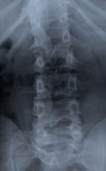
spina bifida
Clinical radiographic examination showing lumbar spinal dysraphism, incomplete ossification of posterior laminar arches, and posterior soft-tissue contouring.
 Zoom
Zoom
spinal dysraphism Meningomyeocele
High-resolution anatomical cross-section demonstrating herniation of the neural placode, nerve roots, and meningeal coverings through the open vertebral defect.
 Zoom
Zoom
Microsurgical Neural Tube Closure & Reconstruction
Operative clinical documentation illustrating delicate microsurgical separation of neural tissue from epithelial placode, untethering of spinal cord, watertight dura-mater reconstruction, and tension-free musculocutaneous flaps.
For consultation and appointment booking, you can reach Dr. Dilip Kiyawat at
-
Phone +91 98220 46043
-
Website drdilipkiyawatneurosurgeon.com