Brain Haemorrhage
BRAIN HEMORRHAGE
Brain haemorrhage, also known as spontaneous intracranial haemorrhage, is a critical medical emergency that occurs when there is bleeding within the brain substance or surrounding meningeal structures without a history of trauma. This condition can have serious consequences; it is a life-threatening event that requires prompt medical and neurosurgical intervention, which is crucial to improving prognosis and saving neurological function.
The commonest cause of spontaneous intracranial haemorrhage is high blood pressure (hypertension), followed by rupture of an intracranial aneurysm, rupture of an arteriovenous malformation (AVM), systemic coagulation defects, and as a side effect of anticoagulant or blood thinner medication consumption.
EMERGENCY HEMATOMA DECOMPRESSION & NEUROLOGICAL SALVAGE
Minimizing intracranial pressure, reversing midline shift, and preserving critical motor and cognitive pathways through timely surgical evacuation
Diagnosis
There is high clinical suspicion when a patient complains of sudden severe headache, vomiting, hemiplegia, or altered consciousness. It is confirmed by emergency CT scan and MRI. Brain angiography (DSA/CTA) is the gold standard to diagnose underlying aneurysm or AVM.
Symptoms
Symptoms include sudden onset of severe headache (‘thunderclap’), vomiting, altered sensorium or loss of consciousness, and paralysis of limbs. Severity varies depending on the size and location of the intracranial haemorrhage.
Surgery
Urgent surgical removal within hours of onset may improve paralysis dramatically. This is achieved via precision stereotactic aspiration through a tiny burr hole or open craniotomy. Patients require structured post-operative neuro-physiotherapy.
Rehabilitation
Ongoing multidisciplinary support for both the individual and their caregivers is vital, combining speech therapy, motor re-training, blood pressure management, and cognitive restoration.
Key Causes of Spontaneous Brain Haemorrhage
Identifying the underlying vascular mechanism is crucial for targeted treatment and preventing re-bleeding:
Hypertension (High BP)
Chronic untreated high blood pressure damages deep perforating cerebral arterioles (Charcot-Bouchard microaneurysms), leading to acute rupture predominantly in the basal ganglia, thalamus, or cerebellum.
Cerebral Aneurysm Rupture
A weakened out-pouching of a brain artery wall that bursts suddenly, resulting in life-threatening subarachnoid or intracerebral hemorrhage accompanied by an explosive thunderclap headache.
Arteriovenous Malformation (AVM)
A congenital tangle of abnormal, high-pressure blood vessels connecting arteries directly to veins without capillary beds, vulnerable to spontaneous rupture in younger individuals.
Coagulation Defects & Blood Thinners
Systemic bleeding diatheses or antiplatelet/anticoagulant medications (warfarin, aspirin, novel oral anticoagulants) that impair normal clot formation following minor vascular insults.
Clinical Radiographs & Burr Hole Aspiration Case
Authentic imaging series demonstrating Dr. Dilip Kiyawat's precision stereotactic blood clot evacuation:
 Zoom
Zoom
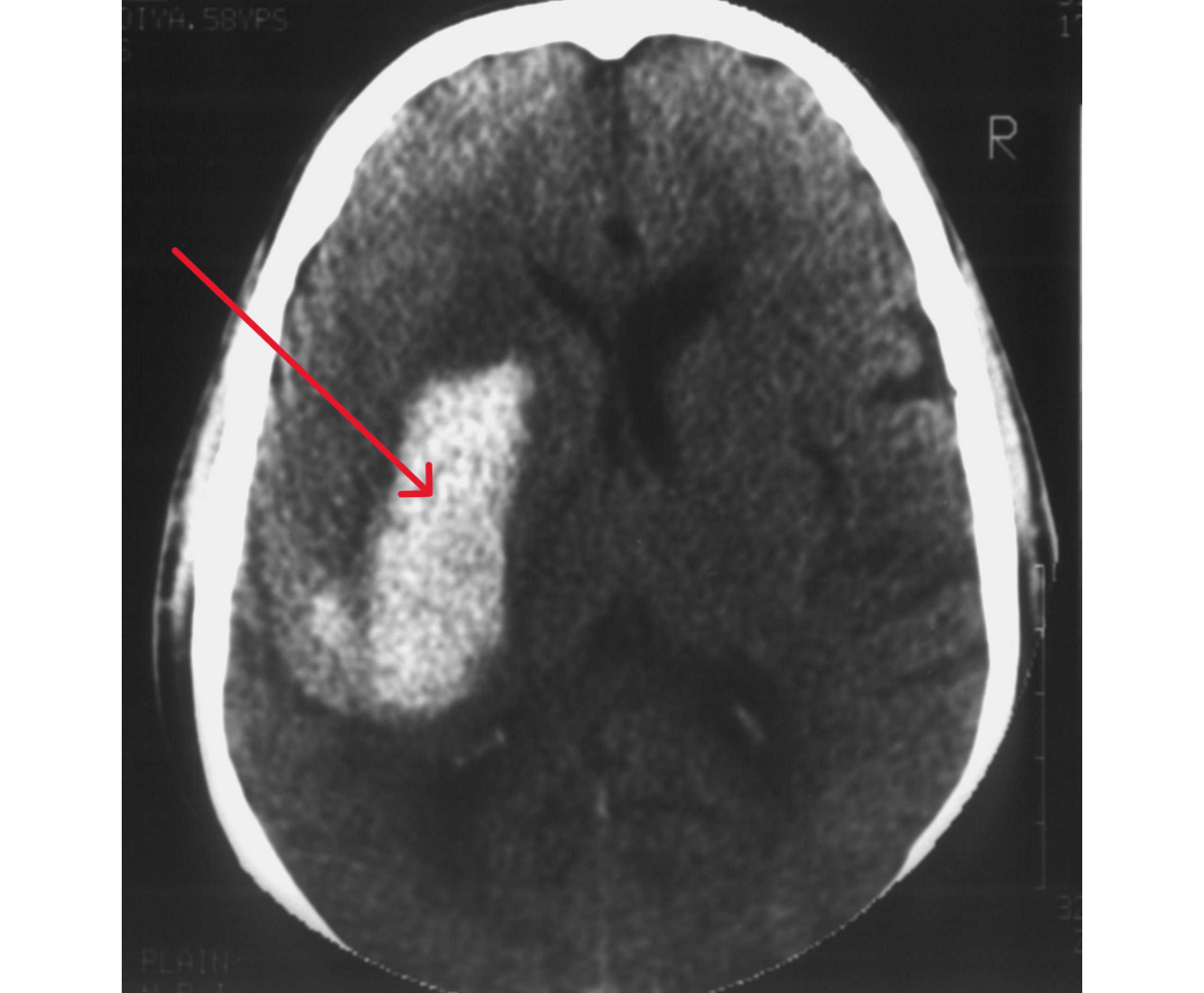
Brain Haemorrhage Noted on Right Side of Brain
Emergency non-contrast CT brain scan identifying acute, dense intracerebral haemorrhage in the right hemisphere with perilesional edema, ventricular compression, and mass effect.
 Zoom
Zoom
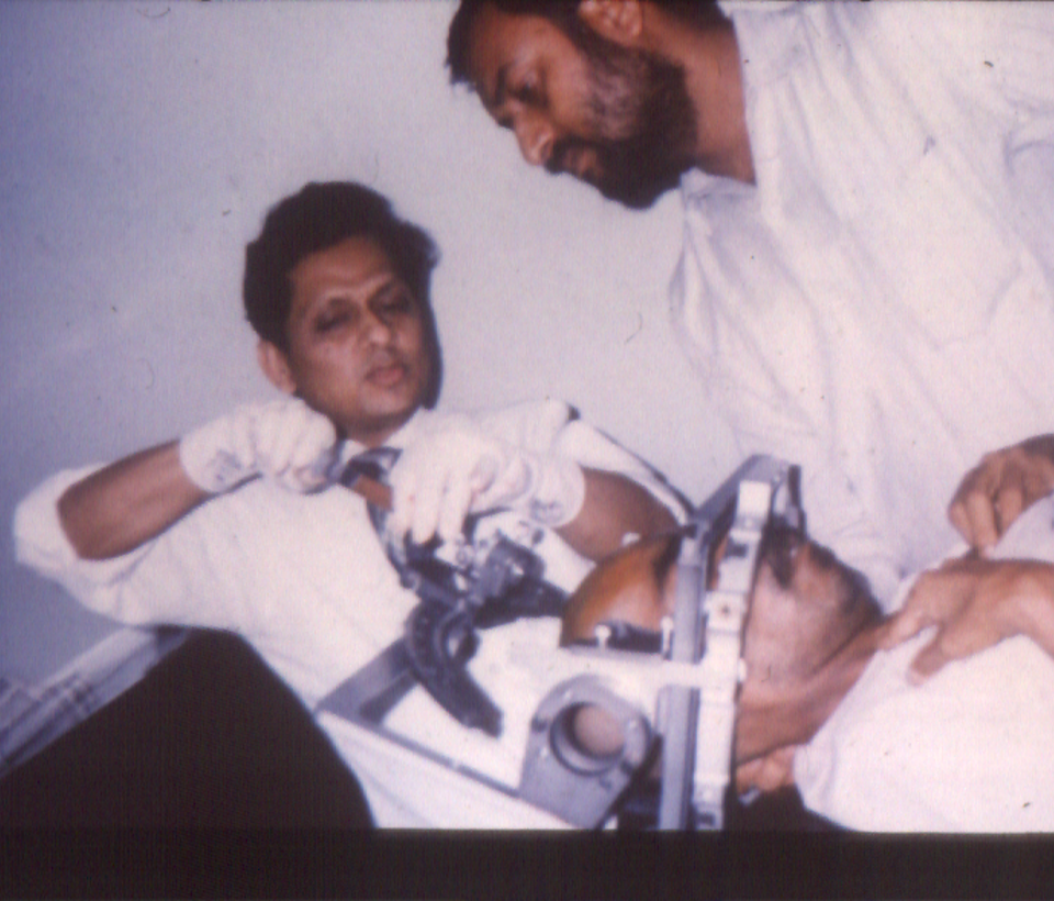
Stereotactic Aspiration Through Tiny Burr Hole
Computer-assisted 3D stereotactic trajectory targeting the core of the hematoma, allowing controlled evacuation of the blood clot through a minimally invasive 18mm burr hole opening.
 Zoom
Zoom
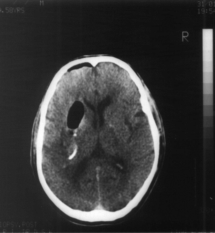
Scan After Aspiration of Blood Clot
Follow-up CT scan confirming successful evacuation of the intracerebral hematoma, decompression of the deep brain structures, and prompt resolution of life-threatening midline shift.
For emergency consultation and appointment booking, you can reach Dr. Dilip Kiyawat at
-
Phone / 24/7 Emergency +91 98220 46043
-
Website drdilipkiyawatneurosurgeon.com