Brain Tumor Surgery
BRAIN TUMOR SURGERY
Brain tumours are abnormal growths of cells within the brain or the surrounding structures. They can be benign (non-cancerous) or malignant (cancerous) and can affect people of all ages. Primary brain tumors are tumors that originate in the brain itself, as opposed to secondary tumors that spread to the brain from other parts of the body.
Various Types of Primary Brain Tumors
MICROSURGICAL RESECTION & NEURONAVIGATION
Achieving safe gross-total tumor excision using high-precision surgical microscopy, intraoperative monitoring, and minimal collateral brain disturbance
Investigations
The presence of a brain tumour is confirmed by high-resolution contrast MRI. It clearly demarcates margins, surrounding brain involvement, and mass effect, guiding a safe and short trajectory to approach the lesion.
Symptoms
Brain tumours may remain silent initially. Common manifestations include progressive morning headaches, vomiting, blurred or double vision, balance instability, new-onset seizures, cognitive changes, and focal limb weakness.
Treatment
The primary treatment for most brain tumors is precise microsurgical resection. The neurosurgeon strives to remove as much of the tumor as possible while scrupulously preserving healthy, eloquent brain parenchyma.
Rehabilitation
Comprehensive post-operative recovery integrates neuro-physiotherapy, speech and occupational therapy, alongside emotional and psychological counseling crucial for patients and their families.
Neurosurgical Procedures to Approach the Brain Tumor
Dr. Dilip Kiyawat employs tailored neurosurgical techniques customized to tumor histology, location, and relationship to vital neural structures:
Craniotomy
In craniotomy, a bone flap is temporarily removed to locate and excise the tumour under microscopic vision. Once the tumour is removed, the bone flap is rigidly reattached with titanium plates and mini-screws. It is the gold-standard procedure for brain tumour surgery.
Endoscopic Surgery
This minimally invasive procedure utilizes a high-definition endoscope with camera and light source to access and resect deep intraventricular or skull-base pituitary tumours through small keyhole openings or transnasal pathways.
Stereotactic Radiosurgery
Advanced non-invasive techniques (including Gamma Knife and CyberKnife) delivering cross-fired, sub-millimeter targeted radiation beams without surgical incisions. Ideal for deep lesions, residual disease, or recurrent tumours.
Burr Hole Evacuation
A small (18–20 mm) precision trephine opening is made in the cranial vault to aspirate cystic tumour fluid, haematomas, or abscess collections rapidly and safely.
Stereotactic Biopsy
3D stereotactic coordinates calculate a computer-guided trajectory through a tiny burr hole. Specialized biopsy forceps sample deep-seated lesions beyond the reach of open craniotomy with minimal risk.
Recovery Profile
Recovery is typically rapid with modern microsurgery. Patients usually emerge from the operating theatre fully conscious. Hospital discharge typically occurs within 5 to 7 days, tailored to individual tumour dynamics.
Complications Encountered During Brain Tumor Surgery
Meticulous neurosurgical preparation and intraoperative neuromonitoring minimize complications:
Infection
Strict laminar-flow theater protocols and prophylactic antibiotics prevent surgical site infections and meningitis.
Bleeding
Microsurgical bipolar electrocautery and hemostatic agents control bleeding, avoiding postoperative haematomas.
Neurological Deficits
Real-time cortical mapping and electrophysiological monitoring protect eloquent speech and motor pathways.
Cerebral Swelling
Intraoperative brain relaxation and perioperative osmotic agents effectively mitigate cerebral edema.
Seizures
Prophylactic antiepileptic medications protect against perioperative seizures and ensure stable neuronal recovery.
Clinical Case Studies & Operative Radiographs
Authentic diagnostic and operative outcome records from Dr. Dilip Kiyawat's brain tumor surgical archives:
 Zoom
Zoom
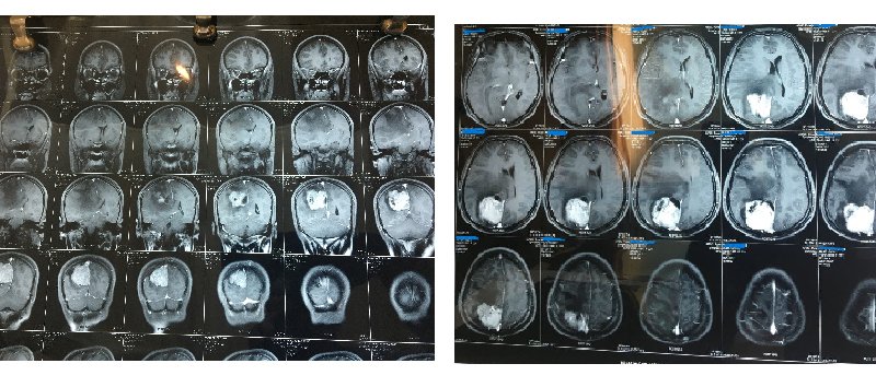
BRAIN TUMOR Before SURGERY
Pre-operative scan illustrating a large intracranial brain tumour causing significant mass effect and brain distortion prior to microsurgical intervention.
 Zoom
Zoom
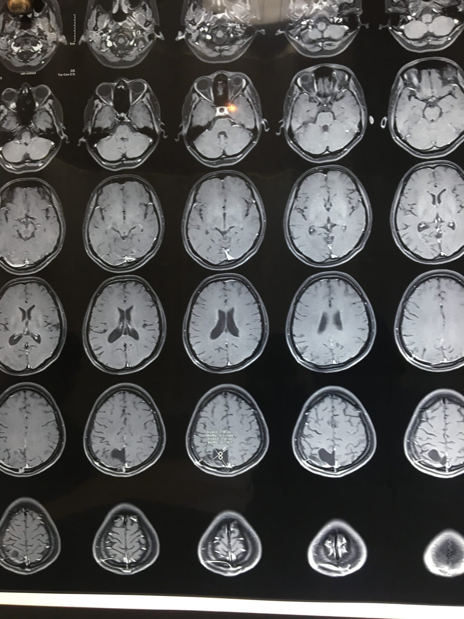
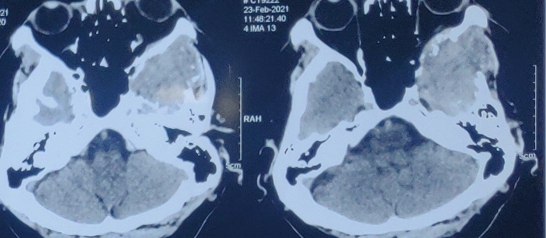
CT Scan After SURGERY
Post-operative follow-up CT scan verifying complete gross-total tumour resection, restoration of midline alignment, and healthy cerebral recovery.
 Zoom
Zoom
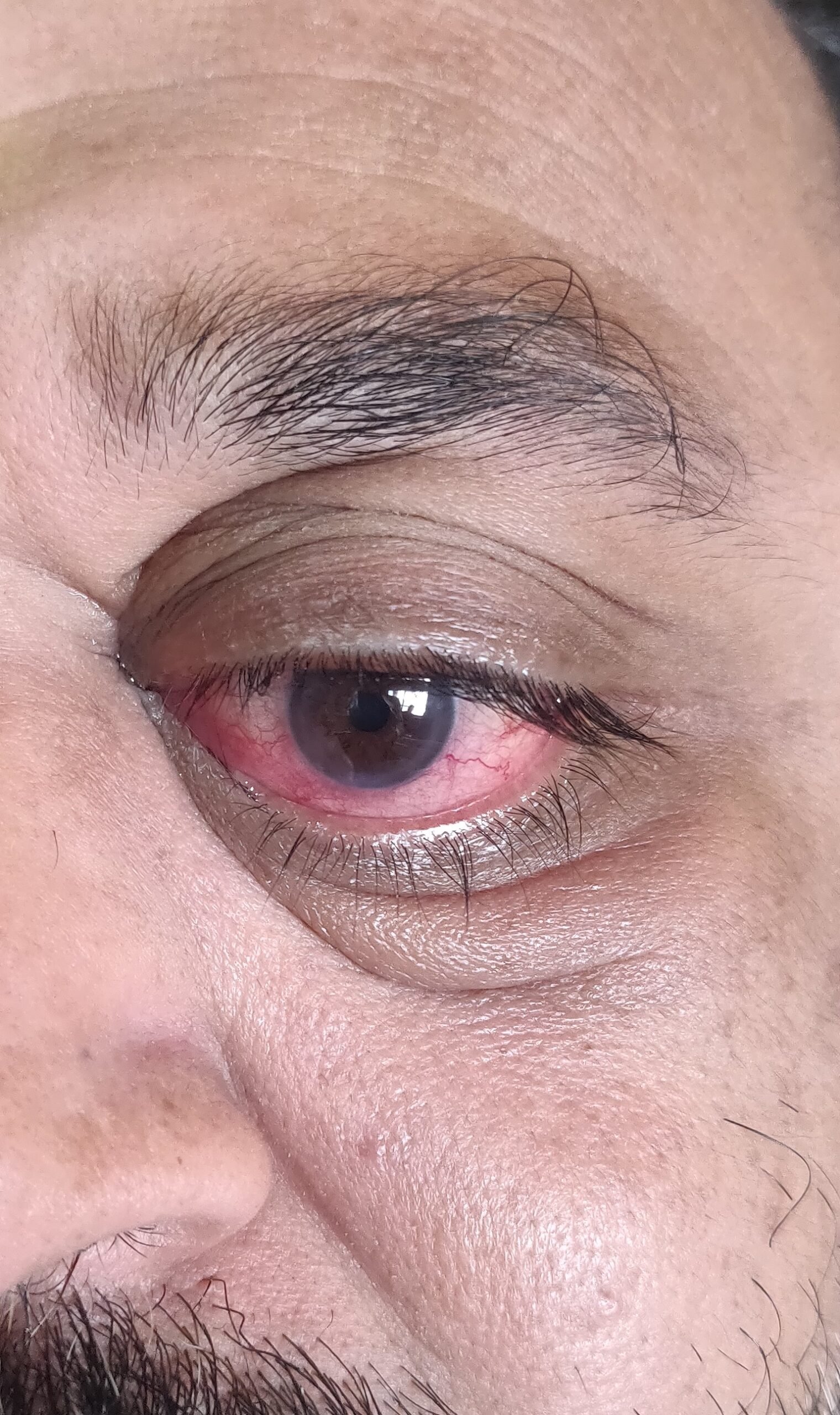
Bulging Eye Due to Retro-Orbital Tumor
Pre-operative clinical photograph showing marked unilateral proptosis resulting from a space-occupying metastatic mass behind the eye.
 Zoom
Zoom
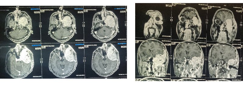
Tumour Behind the Eye (Thyroid Metastasis)
Diagnostic imaging highlighting the retro-bulbar tumour mass compressing the orbital cone, optic nerve, and surrounding ocular structures.
 Zoom
Zoom
Post-Operative Images After Excision
Post-operative radiology demonstrating complete surgical clearance of the retro-orbital metastasis with preservation of vital orbital architecture.
 Zoom
Zoom
After Surgery: Eye Bulge Resolved
Post-operative photograph documenting complete resolution of ocular proptosis, symmetric eye contours, and excellent functional recovery.
For consultation and appointment booking, you can reach Dr. Dilip Kiyawat at
-
Phone +91 98220 46043
-
Website drdilipkiyawatneurosurgeon.com