Cerebral Arteriovenous Malformation (AVM)
CEREBRAL ARTERIOVENOUS MALFORMATION (AVM)
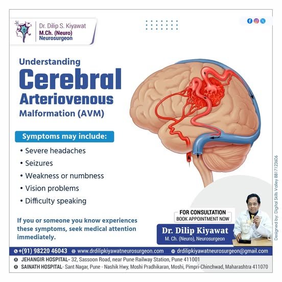
An arteriovenous malformation (AVM) in the brain is an abnormal connection of arteries and veins that disrupts the pattern of normal blood flow and pressure. It is present typically since birth and the exact cause is often unknown. AVMs may remain asymptomatic for many years or they can cause symptoms such as headaches, seizures, or neurological deficits, depending on their size and location. The severity of symptoms often increases if the AVM ruptures, leading to bleeding in the brain.
The diagnosis of AVMs is confirmed through imaging studies such as cerebral angiography, CT scan, or MRI.
Treatment options may include surgical removal through a craniotomy, embolization (blocking blood flow to the AVM), or stereotactic radiosurgery to shrink or obliterate the malformation. This is achieved through a multidisciplinary team of neurosurgeons, interventional radiologists, and neurologists to determine the most appropriate treatment plan based on the specific characteristics of the AVM and the patient’s overall health.
EARLY DIAGNOSIS & MULTIDISCIPLINARY INTERVENTION
Brain AVMs pose a significant lifetime hemorrhage risk if left untreated. Modern neurovascular protocols combine microsurgical resection, targeted endovascular embolization, and stereotactic radiosurgery for definitive vascular obliteration.
Causes
AVMs are often present at birth and result from abnormalities in the development of blood vessels in the brain.
Symptoms
Symptoms can include weakness, numbness, or paralysis in one part of the body, alongside severe headaches.
Treatment
Some small AVMs may be observed without immediate intervention, especially if they are not causing symptoms.
Rehabilitation
Ongoing support for both the individual and their caregivers is crucial for the rehabilitation process and recovery.
Key Insights into Cerebral Arteriovenous Malformations
Understanding presenting neurological symptoms and targeted interventional strategies:
Seizures
A common symptom is seizures, which may be the first sign of an AVM, caused by cortical irritation and vascular steal phenomena altering surrounding blood supply.
Speech and Language Difficulties
AVMs near eloquent speech centers (Broca's or Wernicke's areas) can lead to difficulties in speech fluency, articulation, comprehension, and language processing.
Radiosurgery
Techniques like stereotactic radiosurgery use focused radiation beams to target and treat the AVM nidus over 1–3 years, causing gradual thrombosis and obliteration.
Hemorrhage Risk & Prevention
Rupture leads to life-threatening intracerebral or subarachnoid hemorrhage. Precise Spetzler-Martin grading guides timely treatment to avert catastrophic rebleeding.
Dr. Kiyawat's Multimodal AVM Management Protocol
Comprehensive evaluation and customized intervention tailored to nidus architecture, grade, and eloquent brain localization:
Diagnostic Angiography & Spetzler-Martin Grading
Four-vessel Digital Subtraction Angiography (DSA) and high-resolution MRI evaluate nidus size, depth, arterial feeders, and venous drainage patterns to determine precise surgical risk.
Endovascular Embolization
A neuro-interventional catheter is navigated through the femoral artery into feeding vessels. Liquid embolic agents (Onyx or NBCA glue) block high-pressure feeders, shrinking the nidus.
Microsurgical Resection (Craniotomy)
Through a tailored craniotomy under high-magnification operative microscope, Dr. Kiyawat meticulously isolates, coagulates, and resects the entire tangled nidus while preserving normal cerebral vessels.
Angiographic Confirmation & Neuro-Rehab
Postoperative angiography confirms 100% cure and eradication of the shunt. Structured neuro-rehabilitation aids rapid functional recovery, motor re-training, and speech therapy.
Clinical Radiographs & Cerebral AVM Documentation
Authentic diagnostic radiographs and anatomical documentation from Dr. Dilip Kiyawat's neurovascular archives:
 Zoom
Zoom
CEREBRAL ARTERIO VENOUS MALFORMATION
Authentic diagnostic imaging highlighting the abnormal tangle of dysplastic blood vessels directly shunting arterial blood into the venous circulation.
 Zoom
Zoom
Arteriovenous Malformation Architecture
Comprehensive medical illustration illustrating how arterial feeders deliver high systemic pressure directly into fragile cerebral draining veins.
 Zoom
Zoom
Digital Subtraction Angiogram (DSA)
Gold-standard cerebral angiogram mapping feeding arterial pedicles, the nidus core, and tortuous venous drainage before surgical or endovascular obliteration.
For consultation regarding Cerebral Arteriovenous Malformation (AVM) evaluation or treatment, reach Dr. Dilip Kiyawat at
-
Phone / Clinic Line +91 98220 46043
-
Website drdilipkiyawatneurosurgeon.com