
Microvascular Decompression for Trigeminal Neuralgia
MICROVASCULAR DECOMPRESSION FOR TRIGEMINAL NEURALGIA
Trigeminal neuralgia is a chronic pain condition with episodes of excruciating, sudden, and stabbing pain. Typically, pain occurs on one side of the face, often triggered by simple activities like touching, chewing, brushing teeth, talking, or even a breeze. It lasts for seconds to minutes. Between the two episodes, individuals may be symptom-free. Pain, though short-lived, is so excruciating and intolerable that he or she may think of committing suicide. Hence the condition is infamously also called a “suicidal disease”.
Medical Management: Management includes medications, such as tab. Carbamazepine, Pregabalin, and antidepressants.
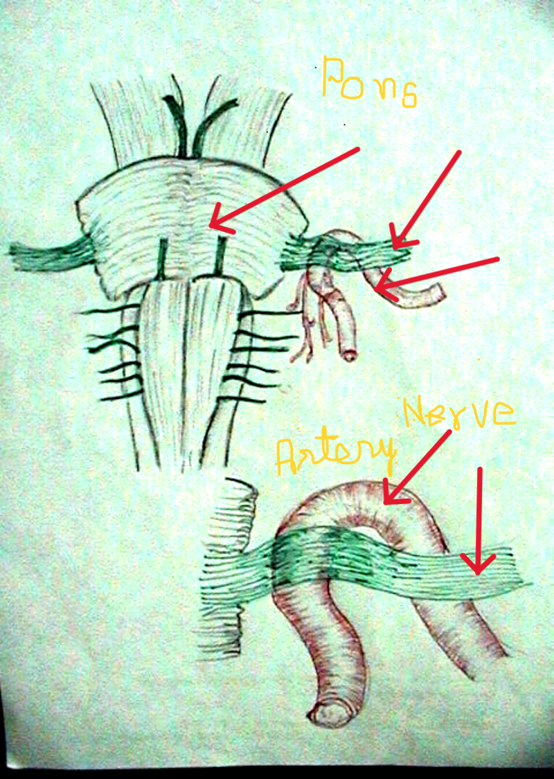
In severe or medication-resistant cases, surgical intervention like Radiofrequency lesions or Microvascular Decompression of the trigeminal nerve (MVD) is done with excellent results. In MVD surgery the trigeminal nerve is exposed near the brain stem where a loop of an artery is found indenting and pulsating on the nerve causing episodic neuralgia.
Once the nerve is relieved, the relief in Trigeminal Neuralgia is dramatic and the patient experiences a new life. Usually the patient is discharged in 3–4 days.
PERMANENT RELIEF WITHOUT FACIAL NUMBNESS
Microvascular decompression cushions the 5th cranial nerve from the pulsating arterial loop with inert Teflon padding, providing immediate pain freedom while preserving complete facial sensation.
Causes
Blood Vessel Compression: The most common cause is the compression of the trigeminal nerve by nearby blood vessels, particularly the superior cerebellar or anterior inferior cerebellar arteries near the brainstem.
Symptoms
Severe Facial Pain: Intense, stabbing, or electric shock-like paroxysmal pain strictly unilateral across the ophthalmic (V1), maxillary (V2), or mandibular (V3) trigeminal divisions.
Treatment
Medical Management with tablet Carbamazepine & Pregabalin is the choice of treatment in early stages. In resistant cases, surgical microvascular decompression (MVD) or radiofrequency rhizotomy is performed.
Rehabilitation & Recovery
Drugs like carbamazepine and oxcarbazepine are gradually tapered off under neurosurgical supervision after MVD, allowing patients to resume normal eating, washing, and speaking comfortably.
Key Insights into Trigeminal Neuralgia
Understanding the neurovascular triggers and secondary compressive etiologies:
Tumor or Cyst Compression
In some cases, benign tumors such as acoustic neuromas, meningiomas, or epidermoid cysts located in the cerebellopontine angle can directly compress the trigeminal root entry zone.
Episodic Trigger Attacks
Pain often occurs in sudden, brief episodes that can be triggered by even mild tactile stimuli such as light touch, a cool breeze, chewing, brushing teeth, smiling, or applying makeup.
Multiple Sclerosis Demyelination
Rarely, demyelination of the central trigeminal nerve root due to multiple sclerosis can lead to trigeminal neuralgia, requiring customized diagnostic MRI sequencing (FIESTA/CISS).
High Long-Term Success Rate
Microvascular decompression provides an exceptional >90% immediate pain cure rate, freeing patients from debilitating pharmaceutical side effects with a fast 3–4 day discharge.
How Microvascular Decompression (MVD) Works
Dr. Dilip Kiyawat's precision microsurgical protocol for permanent cranial nerve protection:
Retrosigmoid Keyhole Exposure
Under general anesthesia, a discreet 3cm incision is made behind the ear. A small keyhole craniotomy (about the size of a postage stamp) provides direct, bloodless access to the cerebellopontine cisterns.
Microscopic Identification of Conflict
Using a high-powered operating microscope, Dr. Kiyawat gently inspects the 5th cranial nerve from the brainstem to Meckel's cave, identifying the precise arterial loop (often the Superior Cerebellar Artery) that is beating against the nerve.
Teflon Felt Cushion Interposition
The offending vessel is mobilized away from the nerve. Microscopic pieces of non-absorbable Teflon sponge are positioned between the vessel and the nerve, permanently dampening pulsatile irritation without injuring nerve fibers.
Immediate Awakening Relief
Because the nerve itself is preserved intact without cutting or burning, the patient wakes up completely free of electric facial shocks while retaining normal tactile sensation in the face, lips, and tongue.
Clinical Radiographs & Trigeminal Neuralgia Cases
Authentic operative documentation and anatomical records from Dr. Dilip Kiyawat's MVD surgical archives:
 Zoom
Zoom
Nerve Compression by an Artery
High-magnification surgical documentation illustrating the arterial loop deeply indenting and pulsating against the root entry zone of the trigeminal nerve before decompression.
 Zoom
Zoom
Microvascular Decompression (MVD)
Clinical schematic showing the permanent interposition of inert Teflon felt, isolating the cranial nerve from arterial pressure waves and curing paroxysmal facial shocks.
 Zoom
Zoom
Trigeminal Nerve Distribution
Detailed neuro-anatomical mapping of the trigeminal sensory dermatomes (V1, V2, V3) and trigger zones, guiding clinical differential diagnosis and targeted surgical planning.
For consultation regarding trigeminal neuralgia or MVD surgery, reach Dr. Dilip Kiyawat at
-
Phone / Clinic Line +91 98220 46043
-
Website drdilipkiyawatneurosurgeon.com